

What Is Oogenesis:
The process of formation of female gametes is called Oogenesis. Oogenesis is a lengthy process. It occurs in ovaries, starts while the female baby is still in the intrauterine stage of development.
The process remains suspended after birth till the onset of puberty. At puberty, it resumes again.
The oogenesis thus can be divided into two stages:
Prenatal stage and Postnatal Stage.
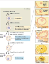
 |
| Complete process of oogenesis in diagram |
Prenatal stage of Oogenesis:
Prenatal stage of oogenesis means oogenesis before birth i.e oogenesis in the intrauterine life. Earlier germ cells called primordial germ cells(primordial means first formed) appear for the first time in the wall of the yolk sac close to the attachment of future umbilical cord. They migrate to the developing ovary by the 4th week of intrauterine life to form oogonia.
In the ovary, they increase in number, by mitosis. Each of them contains 46 single structured chromosomes and 2 N DNA, Single structured chromosome means that it has two arms separated by a centromere1.3. The "N" number refers to the amount of DNA in a cell. (The primordial germ cells and normal somatic cells also contain 16 single chromosomes and 2N amount of DNA.)
The oogonia undergo specialized cell division called meiosis. which is divided into meiosis I and meiosis II . Meiosis l is a reduction division and its characteristic feature is that centromeres of chromo do not split during division. Meiosis ll is a division in which the centromeres do split during division. Meiosis I and meiosis ll both are subdivided into prophase metaphase anaphase and telophase.
The oogonia begin meiosis l by undergoing DNA replication and each of them comes to contain 46 double structured chromosomes and 4N DNA. They are now called primary oocytes.
All the oogonia in both the ovaries are transformed into primary oocyte by the 5th month of intrauterine life and no oogonia are present at birth. Meiosis 1 is prolonged and especially its prophase part.
All primary oocytes remain arrested in prophase from the 5th month of intrauterine life to at least until the onset of puberty. Cell degenerations occur simultaneously and many of primary oocytes become atretic.
The surviving primary oocytes become individually surrounded by a layer of flat epithelial cells derived from the covering mesothelium of the ovary. After birth and until puberty primary oocytes remain dormant. At puberty meiosis 1 is resumed and postnatal part of oogenesis starts.
Postnatal stage of Oogenesis:
At puberty, with the beginning of an ovarian cycle, Postnatal stage of oogenesis starts. With each ovarian cycle, the epithelial cells surrounding each primary oocytes start multiplying. Many layers of cells are thus added around each primary oocyte gradually.
For description, these changes are divided into the following stages.

(a) Primordial follicle:
The primary oocyte together with the surrounding single layer of flat epithelial cells is known as primordial follicle. (Follicle means a small sac). They are the one present principally before birth.

(b) Primary follicle:
Primary oocyte increase in size and the surrounding flat epithelial cells change to cuboidal ones and multiply to produce a stratified epithelium. These cells are now called granulosa cells. The surrounding stromal cells of ovary organize into an inner layer of secretory cells(Theca interna) and an outer layer of connective tissue containing fibroblast-like cells (Theca externa).
Additionally, the granulosa cells and perhaps the oocyte secrete a layer of glycoproteins on the surface of the oocyte (Zona pellucida). This follicle is known as the primary follicle.

(c) Secondary follicle:
As development continues, fluid-filled spaces appear between the granulosa cells. When these spaces coalesce, the antrum is formed and the follicle is termed a secondary follicle.
 |
| Oogenesis |
(d) Tertiary follicle:
Initially, antrum is crescent shaped but with time it greatly enlarges. The granulosa
cells surrounding the primary oocyte remain intact and form cumulus oophorus. At maturity
Roughly in the mid of ovarian cycle, the follicle may be 10 mm or more in diameter. It is called tertiary or Graafian follicle. It may also be called a mature follicle.
As soon as follicle is mature the primary oocyte completes metaphase anaphase and telophase of meiosis 1 to form two daughter cells: secondary oocyte and the first polar body (the latter probably degenerates). Each containing 23 double structured chromosomes and 2N DNA.
The secondary oocyte enters meiosis II in which chromosomes align at the metaphase plate. It is the time when ovulation occurs. After this point oogenesis is completed outside the ovary in the uterine tube where fertilization usually occurs.
The secondary oocyte remains arrested in metaphase of meiosis II until fertilization occurs. At fertilization, secondary oocyte completes metaphase, anaphase, and telophase of meiosis II to form a mature oocyte and the second polar body each containing 23 single structured chromosomes and 1N DNA.
If fertilization does not occur, the secondary oocyte degenerates approximately 24 hours after ovulation. Oogenesis is complete when a mature oocyte with 23 single structured chromosomes with 1N DNA is formed. The second polar body degenerates soon.
Note: The sex chromosomes in primary oocytes are XX. After meiosis 1, the secondary oocyte and the first polar body, each comes to have a double-structured X chromosome because it is a simple reduction division and centromeres do not split. After meiosis ll, the mature oocyte, and the second polar body, each comes to have a single structured X chromosome as it is a simple mitotic division and centromeres split.
 |
| Oogenesis |
You May Also Read It:
- Gametogenesis | Spermatogenesis, And Spermiogenesis
- Cerebral Cortex And Its Division
- Corticospinal Tract Or Pyramidal Tract
- What is cell and Cell Theory
APPROXIMATE NUMBER OF OOCYTES:
A. Primary oocytes:
7 million are present at the 5th month of intrauterine life, 2 million are present at birth and 40000 are present at puberty.
B. Secondary oocytes:
12 are ovulated per year thus up to 480 over the complete reproductive life of
the women (40x12 480).


0 Comments